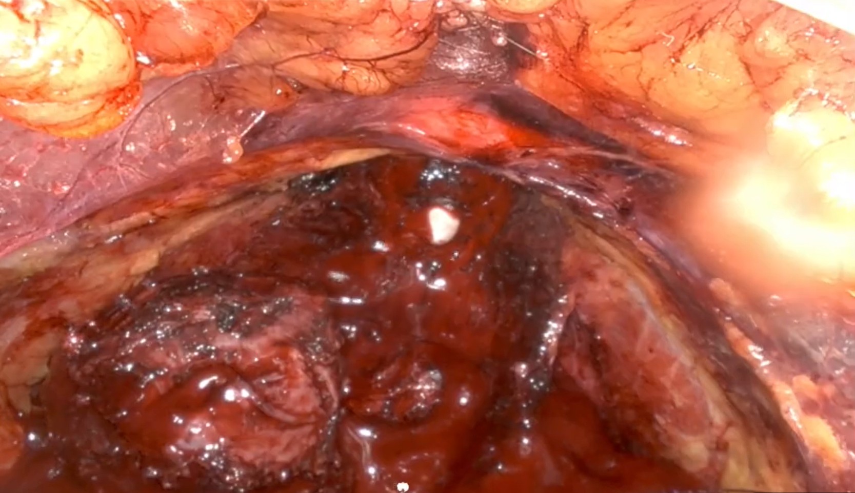
Living prostate tissue at the surgical margin, viewed through the Probentis probe.

Rapidly identify surgical margins at the time and place where it matters the most

A thin optical probe reads the tissue's own biochemical fingerprint, without cutting anything out.

We're seeking to connect with investors and urologists working in prostate cancer care. Reach out and find out how we can work together.

Probentis device is an investigational device and is not yet cleared for sale. Interactions and tissue regions shown are illustrative.

Move the cursor here to find out if and where there is cancer

The Problem

  • 330,000
    New cases of prostate cancer in the US each year1
    Prostate cancer is the leading cancer diagnosis amongst men in the US2
  • Over 100,000
    Radical prostatectomies performed in the US each year3
    Radical prostatectomy is the gold standard surgical treatment for patients with localized prostate cancer4
  • 25% – 30%
    Of patients will have positive surgical margins after surgery5
    The rate and volume of positive surgical margins after radical prostatectomy is one of the highest amongst all cancer surgeries6
  • Urologists cannot visually see the cancer inside the patient during surgery
  • Pre-operative imaging for predicting extracapsular extension can still miss cancer
  • Positive margins are only determined after surgery upon final pathology of the prostate

Our Solution

Leveraging Raman spectroscopy

For instant, in-vivo detection of prostate cancer during surgery

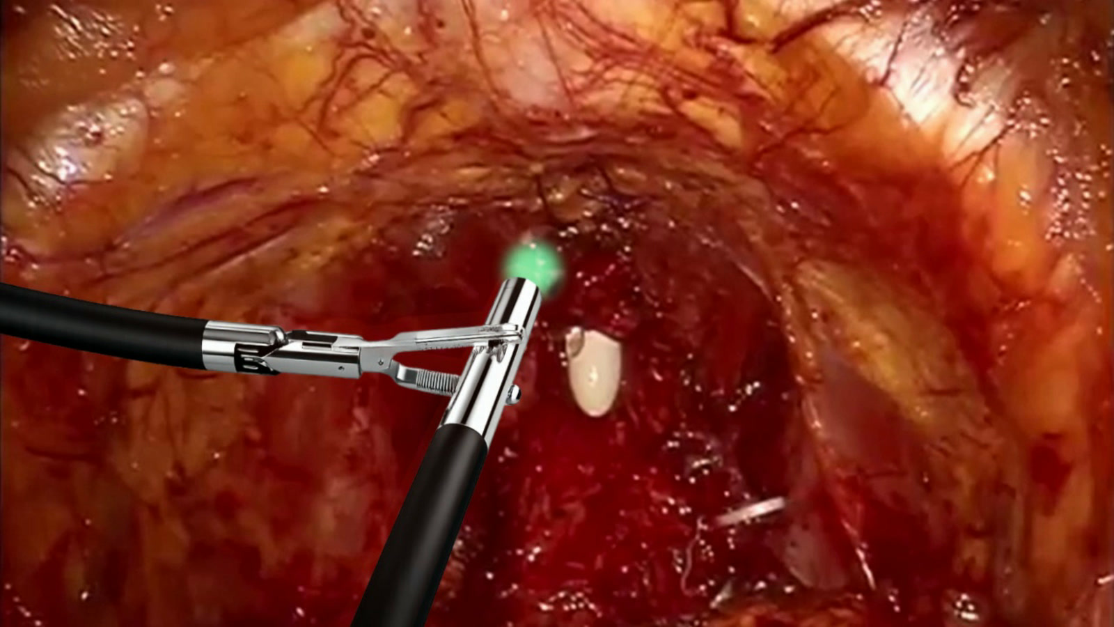
The Probentis probe at the surgical margin, its light green where the reading is clear.
Illustrative. Green marks tissue read as clear.

Highly accurate classification model

Trained and validated on a large dataset of spectra from human prostate tissue, acquired from patients undergoing biopsy

  • 4,110
    spectra acquired
  • 839
    biopsy cores
  • 152
    patients

Model currently based on measurements acquired on freshly biopsied prostate cores (and not on surgical margins). Clinical studies are ongoing.

  • Negative Predictive Value 96.3%
  • Sensitivity 91.0%
  • Specificity 80.0%
  • Accuracy 87.6%
Proprietary dataset. Details of the cohort are available on request.

A "paradigm changing" technology

Integrates with the current workflow

How it works

PROSTATE · CROSS-SECTION suspicious tissue sub-mm Raman probe 785 nm laser Raman spectrum the tissue's biochemical fingerprint Raman shift (cm⁻¹) intensity Tryptophan ~873 Blood ~1248 Proteins ~1650 trained classifier CANCER positive at the margin SAFE clear to retain +ve MALIGNANCY

hover any labelled element for the science →

Publications

Biomedical Optics Express

High-sensitivity Raman spectroscopy for prostate cancer detection and tissue extraction guidance(opens in a new tab)

Dooley MJ, et al.

2026 · 17(7) · 3763–3775

Researchers tested the probe on prostate biopsy tissue from 152 men. It read 3,342 light signatures from 836 samples. The software classified each signature as cancer or clear. A single model caught 80 cancers in every 100. Applying 5 filters in sequence raised that to 90. When the probe called a sample clear, it was right 95 times in 100. The system prioritised catching cancer while clearing 62 in 100 samples without cancer. That cuts the samples needed for diagnosis by almost half. Every reading was taken on tissue already removed from the patient.

Journal of Biophotonics

Raman spectroscopy system for real-time diagnosis of clinically significant prostate cancer tissue(opens in a new tab)

van Breugel SJ, et al.

2023 · 16(5) · e202200334

An earlier study tested the same approach on 84 men. The probe read 2,395 light signatures from 599 biopsy samples. It found clinically significant cancer in 90 of every 100 cases. It correctly cleared about 80 in 100 samples without significant cancer. It also graded the cancers it found. It separated the more aggressive cancers from the less aggressive ones, correctly 90 times in 100. These readings were taken at routine biopsy appointments, on tissue just removed.
Let’s Connect!

Learn what we’re building, or tell us how we could work together.

Contact Us